Probably one of the best events on the topic of no virus in recent history was the court case between Dr. Stefan Lanka and Dr. David Bardens. Jamie went to considerable lengths to dig up and translate the court proceedings in a thread on Twitter that can be reviewed here.
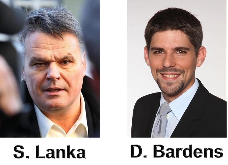
24 November 2011 the German Virologist Dr. Stefan Lanka offered a prize of €100k for a scientific publication in which the alleged existence of the “measles virus” is proven. He did this to raise awareness to what he believed was fraudulent science behind mandatory measles vaccinations.
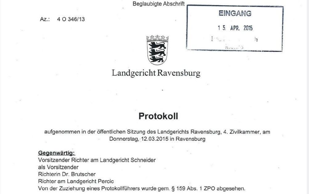
This Challenge was undertaken by Dr. David Bardens who submitted 6 papers he believed proved the existence of the measles virus and took it to Ravensburg Regional Court on non payment. An Ad Hoc judgment was made on 12 March 2015 by Judge Schneider before any rebuttal from Dr. Lanka.
If you search for this court case this is normally what you are met with in the search results. Piles of articles showing that Dr. Lanka lost because of this first court case decision but nothing can be further from the truth.
The Lanka Court Case – Part 1
This Ad Hoc judgment ordered Lanka to pay the prize money to Bardens. Lanka appealed the decision and it was taken to the Stuttgart Higher Regional Court where they would let Lanka make a scientific rebuttal.
The online court records can be reviewed by following the below link:
He was a Bacteriologist with no practical or published competence in the field of virology. His cross examination is recorded in minutes at Ravensburg Court.
It is written in German and a translation app was used, if German speakers could verify the translations that would be of great help. The words of importance are unambiguous but just for the record it is a translated version (all block quoted text in the Lanka court case sections are translations from the court proceedings).
The 6 seminal papers Dr. David Bardens listed as hard concrete evidence that measles virus is causal are:
Enders JF, Peebles TC. Propagation in tissue cultures of cytopathogenic agents from patients with measles. Proc Soc Exp Biol Med. 1954 Jun;86(2):277–286.
Bech V, Magnus Pv. Studies on measles virus in monkey kidney tissue cultures. Acta Pathol Microbiol Scand. 1959; 42(1): 75–85
Horikami SM, Moyer SA. Structure, Transcription, and Replication of measles Virus. Curr Top Microbiol Immunol. 1995; 191: 35–50.
Nakai M, Imagawa DT. Electron microscopy of measels virus replication. J Virol. 1969 Feb; 3(2): 187–97.
Lund GA, Tyrell, DL, Bradley RD, Scraba DG. The molecular length of measles virus RNA and the structural organization of measles nucleocapsids. J Gen Virol. 1984 Sep;65 (Pt 9):1535–42.
Daikoku E, Morita C, Kohno T, Sano K. Analysis of Morphology and Infectivity of measles Virus Particles. Bulletin of the Osaka Medical College. 2007; 53(2): 107–14.
In the court case they focus a lot on the examination of one specific paper by John Enders in 1954. The so called isolation of the measles virus which was coincidently the first time the technique of cell culture isolation was used and is still used for every isolation of a virus in virology.
Explain: The contribution by Enders ^~^ Peebles 1954 definitely fulfils the Henle-Koch postulates of classes formulations No. 1 and 2. There is even a certain biochemical characterization (temperature sensitivity) and a statement of size. In the contribution by Bech ^~^ from Magnus 1958, the third classic Hanle-Koch postulate is also fulfilled. We have additionally demonstrated in this paper the defense reaction which is relevant in the expanded version of these postulates as stated. In fact, however, an experiment in the sense of the 4th classically formulated Henle-Koch postulate was not carried out at that time. As for the other three original papers, these deal significantly with the size and electron microscopic representation of the measles virus and fall out of this review to some extent. The overview article from 1995 then cites and present several articles which, with regard to the measles virus, fulfill all postulates no. 1 to 4 in the classis formulation.
In cross examination of this paper Podbielski makes 2 major admissions:
1. This Paper has “No Negative Control”:
Page 7: I cannot now say whether there is an article that comprehensively presents the same things as the original articles mentioned without showing their methodological weaknesses, for example with the negative controls that are in fact missing. In this context, I would like to point out again that certain parts of the experimental set-up in the original articles from ‘54 and ‘58 do have a certain control function. The following seems decisive to me: Such scientific articles are used for follow-up work by other scientists.
2. It does not fulfill Koch’s Postulates, which are the scientific criteria laid out for proof of existence of a pathogen.
Page 8: When Assessor Schreiner followed up whether this circumstance reduces the evidential value: No, as biological research has been carried out for many decades, this is not the case. When asked by Assessor Schreiner whether the criticism of the early original work, for example that the work from 1954 did not fulfill Henle-Koch’s postulate 3. does not lead to this work being unusable, or whether one can base anything on such work at all: It is not the task of specialist articles on microbiological matters that each specialist article taken by itself immediately contains all four of these Henle-Koch postulates Fulfills; as we can see, some articles do not deal with it at all. Each article has its own scope and work content. If you wanted to comprehensively meet the requirements of all four Henle-Koch postulates in one article, the article would probably be so lengthy that it might not even be suitable for publication in view of the editor’s specifications.
Podbielski attempts to hide the lack of controls in the paper by stating that it is an “old paper” on which to build. This is a major problem as you will see soon. He also tries to glaze over the the fact that it doesn’t fulfill Koch’s Postulates and in a stunning admission none will.
I really don’t know of a single work that, taken by itself, would fulfill all four postulates.
He also lays the foundations for what is used by those who wish to lie about this trial; that they “could” satisfy Koch’s Postulates but it would have to be a very very long paper. This is a made up fluff in an attempt to obscure the zero evidence that he had and the judges agreed.
Each article has its own scope and work content. If you wanted to comprehensively meet the requirements of all four Henle-Koch postulates in one article, the article would probably be so lengthy that it might not even be suitable for publication in view of the editor’s specifications. In and of itself, there is no shortcoming.
To note, this trial wasn’t short of comedy as we see here “expert” Podbielski clashing with Robert Koch Institute Dr. Mankertz disagreeing with each other over whether or not a virus “should” contain a ribosome!
When asked by Assessor Schreiner what the components of the measles virus are, in particular whether the measles virus contains ribosomes: No, the measles virus does not contain any ribonomes. The common definition of the virus is that it has no ribosomes. Assessor Schreiner then addresses the message from the Robert Koch Institute alleged by defendant, according to which the measles virus contains ribosomes: to his question as to whether such a statement would throw the whole concept of measles virus overboard, so to speak: Such a statement would indeed be extremely astonishing, it would attract the greatest attention in the scientific community and could be published with the prospect of great effect.
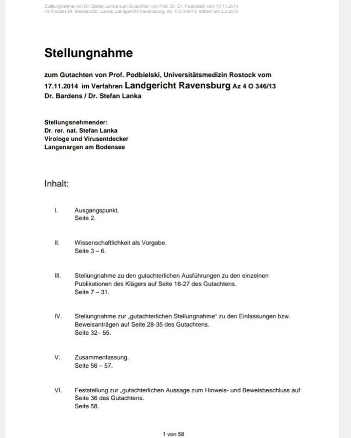
So we move to the Stuttgart Higher Regional Court proceedings where Dr. Lanka produced his 58 page scientific rebuttal. What follows is the core principal behind this rebuttal.
The reason why the trial is heavily focused on Enders 1954 paper is because it is the supposed proof of isolation of the virus. All other papers presented such as genomic sequencing, EM, protein analysis, PCR etc. have to have an isolated virus as a reference, without which it cannot be considered proof.
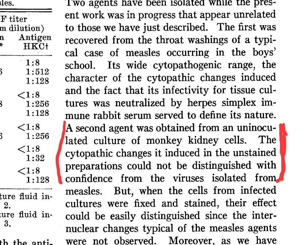
Going back to the comments made by Podbielski of “missing negative control”. This was really only half true. The control was not “missing” it categorically failed. The effects meant to denote the presence of a virus were found in the uninfected sample. The part in the Enders, 1954 paper is shown below and a Twitter thread explaining the control results can be reviewed here.
Explanations of the failed Enders 1954 control are as follows (for those not familiar with it):
Now people (lying shill clowns) who support the Trillion dollar pharma genocide machine like to strawman the second part which reads “they could be differentiated after being fixed and stained” as meaning “fine that is a successful control”. The following has to be considered:
A change (CPE) that is meant to denote the presence of a virus if found, at all, in the control is a failed experiment. The differentiation is not described and irrelevant.
In a court of law this has been described as missing i.e not complete.
Also, Podbielski suggested that Enders is an “old” publication to be built upon and assumes this has been done.
I would like to point out again that certain parts of the experimental set-up in the original articles from ’54 and ’58 do have a certain control function. The fallowing seems decisive to me: Such scientific articles are used for follow-up work by other scientists. As a result, a good cleaning mechanism has been established in the specialist literature, which has recently also affected some articles from top-ranking specialist journals. If the processes presented in the article cannot be reproduced in follow-up experiments, this typically comes to light in articles by other researchers. At least that would have been expected with a topic that has been the subject of such intensive research as measles.
Now this is where it gets interesting as we have clarified. Legally this cell culture technique failed. Unfortunately for virologists and the trillion dollar pharma genocide club, this cell culture technique is the gold standard of every virus isolation since 1954 to present.
Again you will note the comments by Prof Podbielski that “This was an old paper” that science could build on. Well if you know the conclusions to this trial (spoiler alert) you will note; There are no scientific additions with any properly conducted negative controls. As a scientific paper legally requires adequate controls to be performed to be used by government policy. In this case for the measles vaccine, we can only conclude that such a paper does not exist and so we also conclude there is no proof of the existence of any virus by cell culture.
So we fast forward to the closing statements of the unanimous decision of all three judges of the Higher Regional Court of Stuttgart overturning the decision and Granting the plaintiff Dr. Stefan Lanka the Win:
122’ As a result, the appeal was successful, insofar as it is admissible, because the claimant’s criterion of providing evidence of the existence of the measles virus through “a scientific publication” was not met by the plaintiff. Accordingly, the plaintiff is not entitled to any pre-trail attorney’s fees.
123’ 1. The decision on costs is based on §§ 91, 92 Para. 2 No. 1 ZPO
124’ 2. The decision of the provisional enforceability is based on §§ 708 No. 10, 711 ZPO
125’ 3. The revision is not permitted because the requirements of Section 542 (2) ZPO are not met.
Dr. Bardens could then appeal the decision to the Supreme Court of Germany withing a certain period. He decided not to appeal the decision and the time has passed for submission.
Now there are plenty of silly little dim wits out there who believe in the mythical air fairies and big pharma so much that they want to spread the categorical lie that “Lanka won on a technicality, because he said the proof had to be in only 1 paper”. I will show you categorically this is a lie. Yes it stipulates that Lanka wanted “a singular paper” and yes it stipulated that a precise measurement of the virus I.e a characterization of an isolated biological particle, was asked for. Not a drawing which is par for the course in satisfying Kochs Postulates.
Evidence by a single scientific publication 88 The prize money is paid out according to the clear wording of the call for entries 89 if a scientific publication is presented in which the existence of the measles virus is not only claimed, but also proven and its diameter is determined, among other things. The prize money will not be paid if the determination of the diameter of the measles virus is only based on models or drawings like this one.
But the reasons for the judges to accept the singular paper only were based on rational thought not a “technicality” that they didn’t want 100 small letters being “pieced together like a puzzle” as that would not constitute proof. This should be obvious…
92’ Not only the wording speaks for such an understanding, but also the fact that a single work is not only self-contained in terms of its external form and thus clearly delimits the internally structured material, but also that no dispute can arise about through which passage of text which of possibly a large number of works which proof can be provided. With a large number of works that are to be used as proof in their overall view, it can be much more difficult to bring each of the works to a comparable and meaningful level in terms of method and content. In addition, it reduces the effort of the test considerably if the proof has to be provided in a work according to the wording. It is obvious that the defendant, which is also recognizable to third parties, cannot wish for around 50, 100 or 500 different works to be submitted, from which individual text passages or sections are then put together like a puzzle in order to then be able to Reasons of practicability and reasonableness speak in favor of understanding the call for tenders in the way that the wording of them makes a statement in the overall context.
There is also a cry that “because more than one paper was submitted Lanka got off”. This is also a blatant lie spelt out clearly below. There was no limit to the amount of papers you could submit.
Finally, there are no criteria for a meaningful limitation of the number of works to be submitted as evidence in the text of the advert, and such criteria are also not evident: 95’ – Contrary to the regional court – it can also…
But Lanka asked for specific things like “size of the virus”. Obviously if you have an isolated particle you should know its exact size. Problem is that Prof Podbieski noted in his cross examination “he didn’t know but they were all different”.
When asked by Assessor Schreiner how big the measles virus is now: Page 11’ I can’t give any numbers by heart. I have already explained in more detail in my expert report that and why the size information is variable and can be found in the literature discussed.
But onto the most Iron Clad and irrefutable piece of logic that throws the idea of Lanka’s “luck” out of the window. Enders isolation paper “should” have been enough to suffice for proof of existence of the measles virus. One singular paper… The German judicial system disagreed.
The Lanka Court Case – Part 2
In Part 1 we saw how German Virologist Stefan Lanka won his court case showing that there was no proof that the measles virus exists. Really he proved that no virus has ever been isolated as the reason why he won was based entirely on lack of controls.
The isolation method of a supposed virus was dreamt up by John Enders in 1954 who went on to win a Nobel Prize. He took a culture of monkey kidney cells, antibiotics, fetal bovine serum and human samples assumed to contain a virus. He then stresses the culture over days.
When the kidney cells broke down, also called “Cytopathic Effect” (CPE) he pointed at this culture and said “look a virus did that”. The scientifically or logically minded will ask: Was it definitely a virus that did that? How can you tell, you assumed it was there in the first place.
A control is needed to show that it is the variable (virus) causing CPE and not the mixture of other ingredients. So Enders took all those ingredients without adding “infected sample” and still the results showed CPE, meaning it was not something in the human sample causing the effect.
It says that the samples were then distinguishable after being “fixed and stained” but if you are claiming this CPE denotes the presence of a virus and CPE occurred when there could not possibly have been one. Hence the control showed the experiment void.
Bizarrely though, instead of voiding the experiment, the halls of science gave him a Nobel Prize and incorporated his technique into every single experiment “isolating” a virus. This same technique is still being used today and is almost identically to the WHO protocol.
So if we cast our thoughts back to Part 1 in the trial where Podbielski suggests that this “old” technique was presumably built upon since. His assumption was clearly wrong as he was unable to present a single paper with this adequate control showing “something” pathogenic.
I would like to point out again that certain parts of the experimental set-up in the original articles from ’54 and ’58 do have a certain control function. The fallowing seems decisive to me: Such scientific articles are used for follow-up work by other scientists. As a result, a good cleaning mechanism has been established in the specialist literature, which has recently also affected some articles from top-ranking specialist journals. If the processes presented in the article cannot be reproduced in follow-up experiments, this typically comes to light in articles by other researchers. At least that would have been to be expected with a topic that has been the subject of such intensive research as measles.
As part of Stefan Lankas 58 page scientific rebuttal to the Enders paper he instructed a lab to carry out a control experiment, using WHO protocols and materials in a rudimentary test. Here is the description in the court documents and the slides.
The attempt
On behalf of Dr. Lanka verified whether agents other than the alleged measles virus can also lead to cell fusion with resulting cell death (=syncytia formation) in cell cultures that looks exactly like the one in the standardized protocol that, based on the 1954 publication by Enders & Peebles for the Detection of the measles virus” has become globally binding. For this purpose, the protocol of the World Health Organization (WHO) for the detection of measles infection in cell cultures[21] was strictly followed.
The cell lines Vero/CCL-81 and Vero/hSLAM were used. The Vero cells were isolated in March 1962 by Y. Kasumura and Y. Kawakita from the kidney t issues of African monkeys (Cercopithecusaethiops}. They are among the most frequently used continuous mammalian cell lines in research. The Vero/hSLAM cells were transfected with the vector plasmid pCxN2 from Dr. Developed by Yusuke Yanagi. The vector plasmid pCxN2 has a Neomycin resistance gene and an expression plasmid (pCAG-hSLAM) encoding the human signaling lymphocytic activation molecule (hSLAM). The Vero/hSLAM cell line is now recommended for routine ‘isolation’ of the ‘measles virus’. The participants understand isolation as the generation of the effect of syncytial formation in the test tube, which since 1954 has been ad hoc equated with the presence, multiplication and transmission of a “virus” from a person into the test tube, although isolation of a “measles virus” within the meaning of
Lanka’s Latest Control Test
Even though Lanka won the case and had already demonstrated in a court of law that the isolation process was fraudulent he also conducted another control experiment. This time far more comprehensive to squash any doubt. This work was published on 10 March 2022 and the study is discussed below.
Introduction G
Viruses from isolates, eg from bats, are multiplied in cell cultures under harsh culture conditions by giving themby reductionofFetal Calf Serum (FCS) from 10% to 2% or 1% in Dulbecco’s Modified Eagle’s Medium (DMEM) is deprived of a large part of the diet.which conforms to ATCC recommendations. Food deprivation is also routinely combined with high concentrations of Gibco’s triple antibiotics (penicillin/streptomycin antibiotics with amphotericin B antifungal) and sequential blind passage of cell culture supernatants to the next cell culture.[22]
Morphologically, virion amplification leads to cytopathic effects (CPE) that result in cell rounding, ballooning, and cellular degeneration, ultimately manifested by plaque formation in a confluent cell culture. Accordingly, viral particles enriched from these cell culture supernatants can be imaged by electron microscopy. To exclude the hypothesis that harsh stress conditions without virus inoculation might lead to the formation of exosomes[23] that are virion-like, we routinely screened healthy primary human epithelial cells
Subjected to virus amplification protocols. We then isolated total RNA from starved or control cells and supernatants containing viral RNA.
The lab instructed by Lanka strictly followed WHO protocol guidelines to add all of the cell culture ingredients without the possibility of any “virus” being in the culture.
Materials and methods Cell culture
Commercial human primary epithelial cells of passage 3 were thawed and expanded at 4’000 cells/cm2 in 75cm2 flasks at 37°C with 5% CO2 in defined epithelial low calcium medium (without FCS) and 1 x triple antibiotics (Gibco) (control medium, CM).
At >80% confluency, the expansion cells were detached with 5ml Accutase enzyme at 37°C for 1 O minutes. The Accutase was neutralized with 10ml CM, the cells were centrifuged for 5 minutes at 400G, resuspended in 1 ml CM, the living cells were counted using trypan blue staining in the Countess II device (ThermoFisher).
Cells were sawn out for the experiment or parallel rounds of expansion for subsequent experiments. For each experiment, four groups of healthy primary epithelial cells from the same expanded pool were seeded in CM at 4000 cells/ cm2 in 25cm2 flasks and cultured to >50% confluency.
The medium was then replaced with four experimental conditions; for control cells by fresh CM (Control 1) or commercial DMEM supplemented with GlutaMAX, 10% heat-inactivated FCS and 1 x triple antibiotic (Control 2).
Food was withdrawn by replacing CM with DMEM, with 1 % FCS and 3x triple antibiotics, essentially following virion amplification1 protocols (Starvation 1 & 2). The stressed starvation group 2 was additionally treated with 1 O μg total yeast RNA (yRNA) per culture bottle for 1 hour and thoroughly treated with group 1 & 2 before changing the medium washed with phosphate buffered saline (PBS). Two blind passages were then carried out, in which 50% of the supernatant from Starvation groups 1 and 2 was transferred to the next cell culture. The supernatants were cleared of dead cells by centrifugation at 400G for 5 minutes. The control groups received 100% fresh medium.
The experiments were repeated three times in duplicate. The length of the culture under stress defined in the first biological replicate was kept constant for all experiments. No medium change was performed during the stress period.
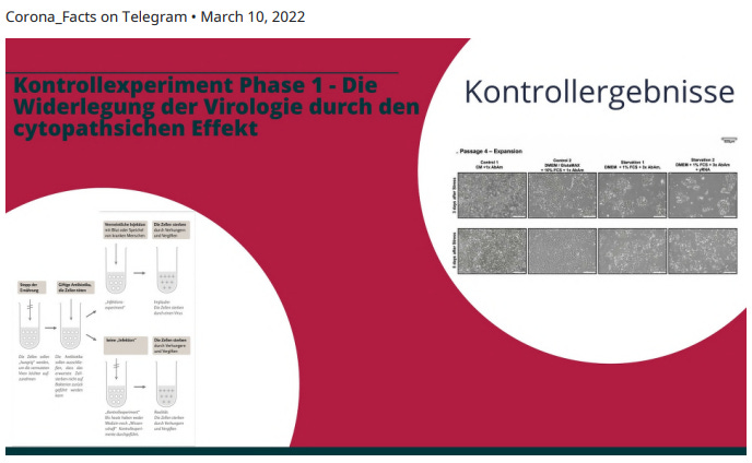
P4: media change in control and stressed cells at about 50% confluency; Control cells cultured to >80% confluency, stressed cells cultured for 5 days after media change.
P5: Media change for control and stressed cells >50% confluency, control cells cultured to >80% confluency, stressed cells cultured for 8 days after media change.
P6/RNA isolation: media change in control and stressed cells at about 50% confluency; Control cells cultured to >80% confluency, stressed cells cultured for 5 days after media change. P6/Crystal violet: media change in control and and stressed cells at 100% confluency; Stress induction for 3 days. A representative photograph of all cell cultures was taken daily at room temperature using a Nikon Eclipse TS100 bright field microscope with a Nikon 1J5 camera, a Nikon FT1 adapter and a 4x objective.
The results are shown below. You can clearly see that as the amounts of antibiotics, removal of nutrients and time increases the cell’s that clearly clump together dying off… Cytopathic Effect. The concentrations of these materials and methods were all done to standard WHO procedure.
And here are the cells “fixed and stained” purple. They do look different and you can “tell the difference between stained and unstained” but that doesn’t change the fact that CPE occurred in the control. Hence proving the cell culture method fraudulent.
Below is a description of the results.
Results
Healthy, primary human epithelial cells were grown over four passages (P3-P6) under optimal culture conditions in defined epithelial control medium with 1 x triple antibiotics (CM).
After the first passage, the cell pool was divided into four groups.
After 3 days in CM, cultures were transferred to either fresh CM (CM, Control 1 ), DMEM/GlutaMAX with 10% FCS, 1 x triple antibiotics (Control 2), or stress medium (Starvation 1 & 2).
During the first stress treatment, the stress medium contained OM EM, 1 % FCS and 3x trip le antibiotics.
The second and third passages were “blind” passages in which 50% of the culture supernatant from the last passage was transferred to the next passage in DMEM, 1 % FCS and 3x triple antibiotics.
The second stress group was additionally treated at each passage with total yeast RNA (yRNA) for one hour before adding the stress medium (Starvation 2).
After transfer to DMEM with 10% FCS, the epithelial cells assumed a flatter morphology than in CM and formed a continuous sheet of cells, which is attributed to the high calcium concentrations in DMEM.
Otherwise, the cells continued to divide normally (Figure 1 A – see below).
In contrast, the cell layers in the stress media shrank to small islands with reduced growth and incipient cell degeneration. During the next two passages, cells incubated with the supernatant of the stressed cells from the previous passage showed increasing CPE with cell-free areas resembling virion-related plaques in the cell sheet, and more dead cells floating in the supernatant (Figure 1 B – see further down).
Confluent cultures under stress (Figure 1 C – see below) stained with crystal violet (Figure 1 D – see below) confirm the pronounced CPE.
Pyknotic cells with condensed nuclei or ballooning cells were predominantly present in the Starvation 1 group and areas of total cell dest ruction or plaques were also observed in the Starvation 1 but predominantly in the Starvation 2 group.
The experiments were performed in three biological replicates and two technical duplicates. All cultures were inspected blindly, with stressed cultures easily identified by drastic changes in morphology.
After three passages, the RNA from the control 1 and the two stressed cell groups and supernatants was isolated using viral RNA kits or TRizol and subjected to nextgeneration sequencing. The amount of total RNA isolated was most abundant in control group 1 (Table 1 – see below) and was of good quality in all groups (data not shown). Further supernatants were further used for the analysis of extracellular particles. The experiments are in progress.
Conclusion
Here is the link to the paper to read the control test section of the Enders 1954 paper. This method is still used today in nearly all isolation studies of viruses… Despite it being proven not to work when Enders designed it in 1954 which is explained in his own paper.
Here is a short video of Lanka summarizing all of this work.
This work was done by Jamie Andrews and a link to his twitter account has been provided in the article. It has been published in dpl’s substack but under a separate newsletter created for Jamie’s work. It has been published here with the approval by the author.