Probably one of the best events on the topic of no virus in recent history was the court case between Dr. Stefan Lanka and Dr. David Bardens. Jamie went to considerable lengths to dig up and translate the court proceedings in a thread on Twitter that can be reviewed here.
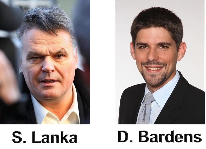
24 November 2011 the German Virologist Dr. Stefan Lanka offered a prize of €100k for a scientific publication in which the alleged existence of the “measles virus” is proven. He did this to raise awareness to what he believed was fraudulent science behind mandatory measles vaccinations.
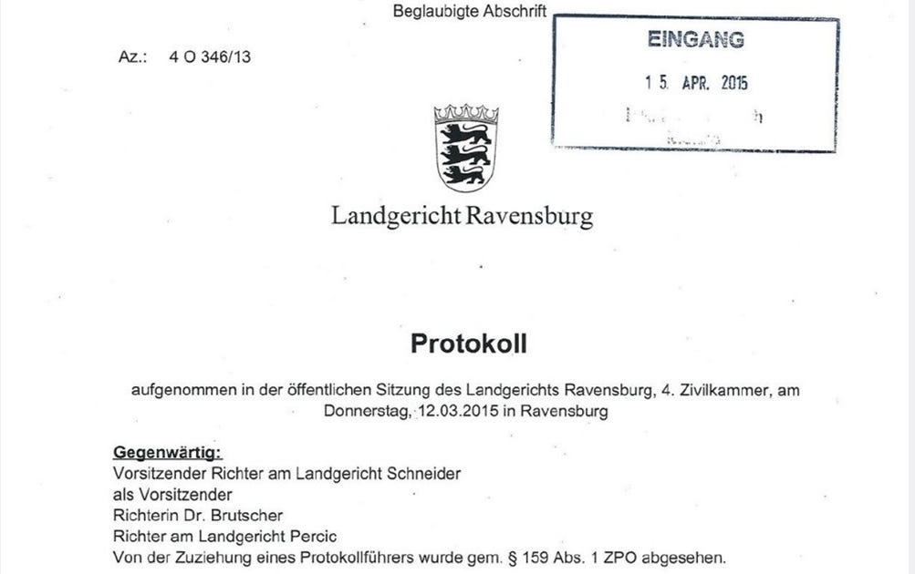
This Challenge was undertaken by Dr. David Bardens who submitted 6 papers he believed proved the existence of the measles virus and took it to Ravensburg Regional Court on non payment. An Ad Hoc judgment was made on 12 March 2015 by Judge Schneider before any rebuttal from Dr. Lanka.
If you search for this court case this is normally what you are met with in the search results. Piles of articles showing that Dr. Lanka lost because of this first court case decision but nothing can be further from the truth.
The Lanka Court Case – Part 1
This Ad Hoc judgment ordered Lanka to pay the prize money to Bardens. Lanka appealed the decision and it was taken to the Stuttgart Higher Regional Court where they would let Lanka make a scientific rebuttal.
The online court records can be reviewed by following the below link:
He was a Bacteriologist with no practical or published competence in the field of virology. His cross examination is recorded in minutes at Ravensburg Court.
It is written in German and a translation app was used, if German speakers could verify the translations that would be of great help. The words of importance are unambiguous but just for the record it is a translated version (all block quoted text in the Lanka court case sections are translations from the court proceedings).
The 6 seminal papers Dr. David Bardens listed as hard concrete evidence that measles virus is causal are:
Enders JF, Peebles TC. Propagation in tissue cultures of cytopathogenic agents from patients with measles. Proc Soc Exp Biol Med. 1954 Jun;86(2):277–286.
Bech V, Magnus Pv. Studies on measles virus in monkey kidney tissue cultures. Acta Pathol Microbiol Scand. 1959; 42(1): 75–85
Horikami SM, Moyer SA. Structure, Transcription, and Replication of measles Virus. Curr Top Microbiol Immunol. 1995; 191: 35–50.
Nakai M, Imagawa DT. Electron microscopy of measels virus replication. J Virol. 1969 Feb; 3(2): 187–97.
Lund GA, Tyrell, DL, Bradley RD, Scraba DG. The molecular length of measles virus RNA and the structural organization of measles nucleocapsids. J Gen Virol. 1984 Sep;65 (Pt 9):1535–42.
Daikoku E, Morita C, Kohno T, Sano K. Analysis of Morphology and Infectivity of measles Virus Particles. Bulletin of the Osaka Medical College. 2007; 53(2): 107–14.
In the court case they focus a lot on the examination of one specific paper by John Enders in 1954. The so called isolation of the measles virus which was coincidently the first time the technique of cell culture isolation was used and is still used for every isolation of a virus in virology.
Explain: The contribution by Enders ^~^ Peebles 1954 definitely fulfils the Henle-Koch postulates of classes formulations No. 1 and 2. There is even a certain biochemical characterization (temperature sensitivity) and a statement of size. In the contribution by Bech ^~^ from Magnus 1958, the third classic Hanle-Koch postulate is also fulfilled. We have additionally demonstrated in this paper the defense reaction which is relevant in the expanded version of these postulates as stated. In fact, however, an experiment in the sense of the 4th classically formulated Henle-Koch postulate was not carried out at that time. As for the other three original papers, these deal significantly with the size and electron microscopic representation of the measles virus and fall out of this review to some extent. The overview article from 1995 then cites and present several articles which, with regard to the measles virus, fulfill all postulates no. 1 to 4 in the classis formulation.
In cross examination of this paper Podbielski makes 2 major admissions:
1. This Paper has “No Negative Control”:
Page 7: I cannot now say whether there is an article that comprehensively presents the same things as the original articles mentioned without showing their methodological weaknesses, for example with the negative controls that are in fact missing. In this context, I would like to point out again that certain parts of the experimental set-up in the original articles from ‘54 and ‘58 do have a certain control function. The following seems decisive to me: Such scientific articles are used for follow-up work by other scientists.
2. It does not fulfill Koch’s Postulates, which are the scientific criteria laid out for proof of existence of a pathogen.
Page 8: When Assessor Schreiner followed up whether this circumstance reduces the evidential value: No, as biological research has been carried out for many decades, this is not the case. When asked by Assessor Schreiner whether the criticism of the early original work, for example that the work from 1954 did not fulfill Henle-Koch’s postulate 3. does not lead to this work being unusable, or whether one can base anything on such work at all: It is not the task of specialist articles on microbiological matters that each specialist article taken by itself immediately contains all four of these Henle-Koch postulates Fulfills; as we can see, some articles do not deal with it at all. Each article has its own scope and work content. If you wanted to comprehensively meet the requirements of all four Henle-Koch postulates in one article, the article would probably be so lengthy that it might not even be suitable for publication in view of the editor’s specifications.
Podbielski attempts to hide the lack of controls in the paper by stating that it is an “old paper” on which to build. This is a major problem as you will see soon. He also tries to glaze over the the fact that it doesn’t fulfill Koch’s Postulates and in a stunning admission none will.
I really don’t know of a single work that, taken by itself, would fulfill all four postulates.
He also lays the foundations for what is used by those who wish to lie about this trial; that they “could” satisfy Koch’s Postulates but it would have to be a very very long paper. This is a made up fluff in an attempt to obscure the zero evidence that he had and the judges agreed.
Each article has its own scope and work content. If you wanted to comprehensively meet the requirements of all four Henle-Koch postulates in one article, the article would probably be so lengthy that it might not even be suitable for publication in view of the editor’s specifications. In and of itself, there is no shortcoming.
To note, this trial wasn’t short of comedy as we see here “expert” Podbielski clashing with Robert Koch Institute Dr. Mankertz disagreeing with each other over whether or not a virus “should” contain a ribosome!
When asked by Assessor Schreiner what the components of the measles virus are, in particular whether the measles virus contains ribosomes: No, the measles virus does not contain any ribonomes. The common definition of the virus is that it has no ribosomes. Assessor Schreiner then addresses the message from the Robert Koch Institute alleged by defendant, according to which the measles virus contains ribosomes: to his question as to whether such a statement would throw the whole concept of measles virus overboard, so to speak: Such a statement would indeed be extremely astonishing, it would attract the greatest attention in the scientific community and could be published with the prospect of great effect.
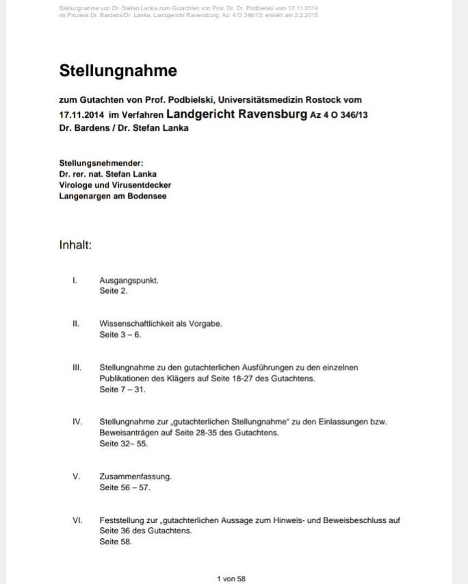
So we move to the Stuttgart Higher Regional Court proceedings where Dr. Lanka produced his 58 page scientific rebuttal. What follows is the core principal behind this rebuttal.
The reason why the trial is heavily focused on Enders 1954 paper is because it is the supposed proof of isolation of the virus. All other papers presented such as genomic sequencing, EM, protein analysis, PCR etc. have to have an isolated virus as a reference, without which it cannot be considered proof.
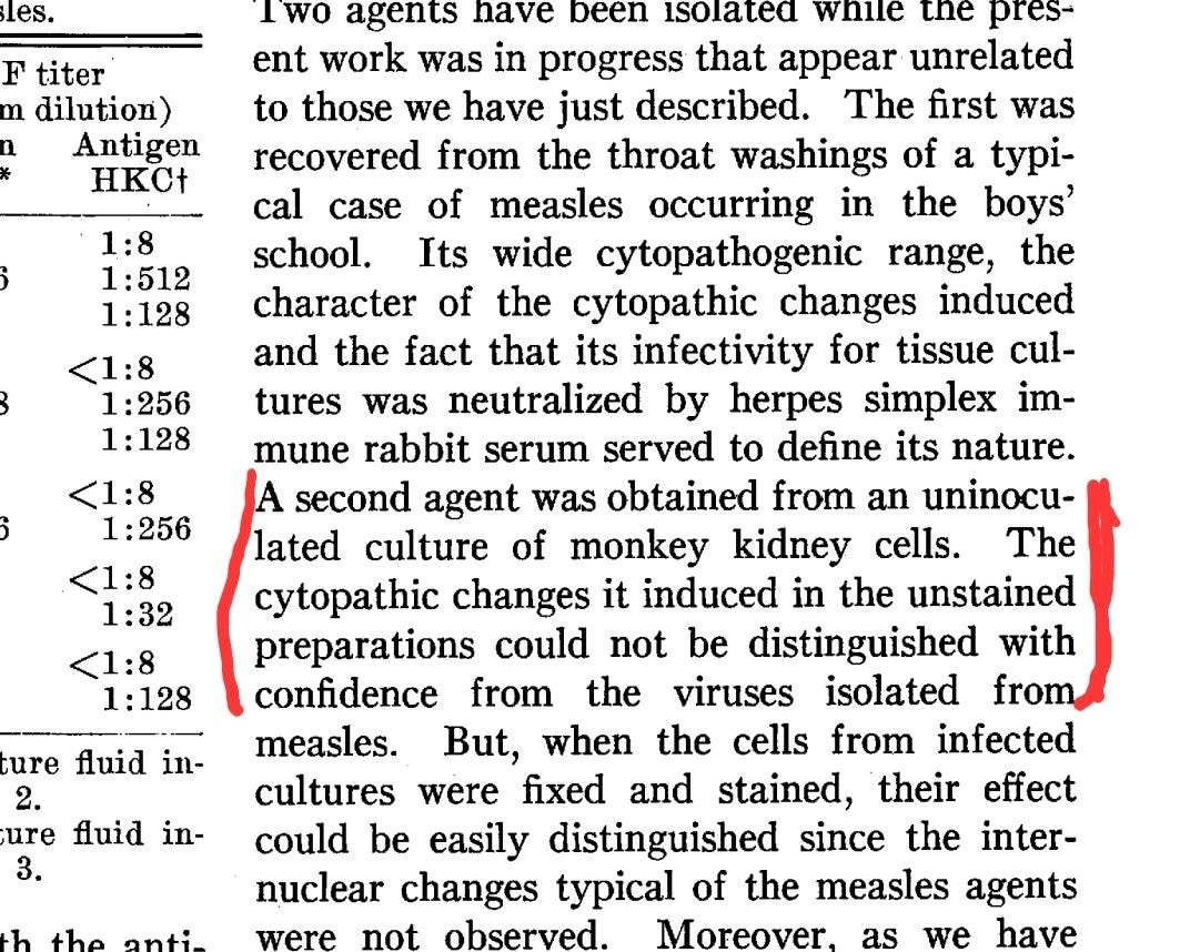
Going back to the comments made by Podbielski of “missing negative control”. This was really only half true. The control was not “missing” it categorically failed. The effects meant to denote the presence of a virus were found in the uninfected sample. The part in the Enders, 1954 paper is shown below and a Twitter thread explaining the control results can be reviewed here.
Explanations of the failed Enders 1954 control are as follows (for those not familiar with it):
Now people (lying shill clowns) who support the Trillion dollar pharma genocide machine like to strawman the second part which reads “they could be differentiated after being fixed and stained” as meaning “fine that is a successful control”. The following has to be considered:
A change (CPE) that is meant to denote the presence of a virus if found, at all, in the control is a failed experiment. The differentiation is not described and irrelevant.
In a court of law this has been described as missing i.e not complete.
Also, Podbielski suggested that Enders is an “old” publication to be built upon and assumes this has been done.
I would like to point out again that certain parts of the experimental set-up in the original articles from ’54 and ’58 do have a certain control function. The fallowing seems decisive to me: Such scientific articles are used for follow-up work by other scientists. As a result, a good cleaning mechanism has been established in the specialist literature, which has recently also affected some articles from top-ranking specialist journals. If the processes presented in the article cannot be reproduced in follow-up experiments, this typically comes to light in articles by other researchers. At least that would have been expected with a topic that has been the subject of such intensive research as measles.
Now this is where it gets interesting as we have clarified. Legally this cell culture technique failed. Unfortunately for virologists and the trillion dollar pharma genocide club, this cell culture technique is the gold standard of every virus isolation since 1954 to present.
Again you will note the comments by Prof Podbielski that “This was an old paper” that science could build on. Well if you know the conclusions to this trial (spoiler alert) you will note; There are no scientific additions with any properly conducted negative controls. As a scientific paper legally requires adequate controls to be performed to be used by government policy. In this case for the measles vaccine, we can only conclude that such a paper does not exist and so we also conclude there is no proof of the existence of any virus by cell culture.
So we fast forward to the closing statements of the unanimous decision of all three judges of the Higher Regional Court of Stuttgart overturning the decision and Granting the plaintiff Dr. Stefan Lanka the Win:
122’ As a result, the appeal was successful, insofar as it is admissible, because the claimant’s criterion of providing evidence of the existence of the measles virus through “a scientific publication” was not met by the plaintiff. Accordingly, the plaintiff is not entitled to any pre-trail attorney’s fees.
123’ 1. The decision on costs is based on §§ 91, 92 Para. 2 No. 1 ZPO
124’ 2. The decision of the provisional enforceability is based on §§ 708 No. 10, 711 ZPO
125’ 3. The revision is not permitted because the requirements of Section 542 (2) ZPO are not met.
Dr. Bardens could then appeal the decision to the Supreme Court of Germany withing a certain period. He decided not to appeal the decision and the time has passed for submission.
Now there are plenty of silly little dim wits out there who believe in the mythical air fairies and big pharma so much that they want to spread the categorical lie that “Lanka won on a technicality, because he said the proof had to be in only 1 paper”. I will show you categorically this is a lie. Yes it stipulates that Lanka wanted “a singular paper” and yes it stipulated that a precise measurement of the virus I.e a characterization of an isolated biological particle, was asked for. Not a drawing which is par for the course in satisfying Kochs Postulates.
Evidence by a single scientific publication 88 The prize money is paid out according to the clear wording of the call for entries 89 if a scientific publication is presented in which the existence of the measles virus is not only claimed, but also proven and its diameter is determined, among other things. The prize money will not be paid if the determination of the diameter of the measles virus is only based on models or drawings like this one.
But the reasons for the judges to accept the singular paper only were based on rational thought not a “technicality” that they didn’t want 100 small letters being “pieced together like a puzzle” as that would not constitute proof. This should be obvious…
92’ Not only the wording speaks for such an understanding, but also the fact that a single work is not only self-contained in terms of its external form and thus clearly delimits the internally structured material, but also that no dispute can arise about through which passage of text which of possibly a large number of works which proof can be provided. With a large number of works that are to be used as proof in their overall view, it can be much more difficult to bring each of the works to a comparable and meaningful level in terms of method and content. In addition, it reduces the effort of the test considerably if the proof has to be provided in a work according to the wording. It is obvious that the defendant, which is also recognizable to third parties, cannot wish for around 50, 100 or 500 different works to be submitted, from which individual text passages or sections are then put together like a puzzle in order to then be able to Reasons of practicability and reasonableness speak in favor of understanding the call for tenders in the way that the wording of them makes a statement in the overall context.
There is also a cry that “because more than one paper was submitted Lanka got off”. This is also a blatant lie spelt out clearly below. There was no limit to the amount of papers you could submit.
Finally, there are no criteria for a meaningful limitation of the number of works to be submitted as evidence in the text of the advert, and such criteria are also not evident: 95’ – Contrary to the regional court – it can also…
But Lanka asked for specific things like “size of the virus”. Obviously if you have an isolated particle you should know its exact size. Problem is that Prof Podbieski noted in his cross examination “he didn’t know but they were all different”.
When asked by Assessor Schreiner how big the measles virus is now: Page 11’ I can’t give any numbers by heart. I have already explained in more detail in my expert report that and why the size information is variable and can be found in the literature discussed.
But onto the most Iron Clad and irrefutable piece of logic that throws the idea of Lanka’s “luck” out of the window. Enders isolation paper “should” have been enough to suffice for proof of existence of the measles virus. One singular paper… The German judicial system disagreed.
The Lanka Court Case – Part 2
In Part 1 we saw how German Virologist Stefan Lanka won his court case showing that there was no proof that the measles virus exists. Really he proved that no virus has ever been isolated as the reason why he won was based entirely on lack of controls.
The isolation method of a supposed virus was dreamt up by John Enders in 1954 who went on to win a Nobel Prize. He took a culture of monkey kidney cells, antibiotics, fetal bovine serum and human samples assumed to contain a virus. He then stresses the culture over days.
When the kidney cells broke down, also called “Cytopathic Effect” (CPE) he pointed at this culture and said “look a virus did that”. The scientifically or logically minded will ask: Was it definitely a virus that did that? How can you tell, you assumed it was there in the first place.
A control is needed to show that it is the variable (virus) causing CPE and not the mixture of other ingredients. So Enders took all those ingredients without adding “infected sample” and still the results showed CPE, meaning it was not something in the human sample causing the effect.
It says that the samples were then distinguishable after being “fixed and stained” but if you are claiming this CPE denotes the presence of a virus and CPE occurred when there could not possibly have been one. Hence the control showed the experiment void.
Bizarrely though, instead of voiding the experiment, the halls of science gave him a Nobel Prize and incorporated his technique into every single experiment “isolating” a virus. This same technique is still being used today and is almost identically to the WHO protocol.
So if we cast our thoughts back to Part 1 in the trial where Podbielski suggests that this “old” technique was presumably built upon since. His assumption was clearly wrong as he was unable to present a single paper with this adequate control showing “something” pathogenic.
I would like to point out again that certain parts of the experimental set-up in the original articles from ’54 and ’58 do have a certain control function. The fallowing seems decisive to me: Such scientific articles are used for follow-up work by other scientists. As a result, a good cleaning mechanism has been established in the specialist literature, which has recently also affected some articles from top-ranking specialist journals. If the processes presented in the article cannot be reproduced in follow-up experiments, this typically comes to light in articles by other researchers. At least that would have been to be expected with a topic that has been the subject of such intensive research as measles.
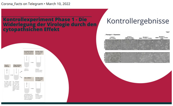
As part of Stefan Lankas 58 page scientific rebuttal to the Enders paper he instructed a lab to carry out a control experiment, using WHO protocols and materials in a rudimentary test. Here is the description in the court documents and the slides.
The attempt
On behalf of Dr. Lanka verified whether agents other than the alleged measles virus can also lead to cell fusion with resulting cell death (=syncytia formation) in cell cultures that looks exactly like the one in the standardized protocol that, based on the 1954 publication by Enders & Peebles for the Detection of the measles virus” has become globally binding. For this purpose, the protocol of the World Health Organization (WHO) for the detection of measles infection in cell cultures[21] was strictly followed.
The cell lines Vero/CCL-81 and Vero/hSLAM were used. The Vero cells were isolated in March 1962 by Y. Kasumura and Y. Kawakita from the kidney t issues of African monkeys (Cercopithecusaethiops}. They are among the most frequently used continuous mammalian cell lines in research. The Vero/hSLAM cells were transfected with the vector plasmid pCxN2 from Dr. Developed by Yusuke Yanagi. The vector plasmid pCxN2 has a Neomycin resistance gene and an expression plasmid (pCAG-hSLAM) encoding the human signaling lymphocytic activation molecule (hSLAM). The Vero/hSLAM cell line is now recommended for routine ‘isolation’ of the ‘measles virus’. The participants understand isolation as the generation of the effect of syncytial formation in the test tube, which since 1954 has been ad hoc equated with the presence, multiplication and transmission of a “virus” from a person into the test tube, although isolation of a “measles virus” within the meaning of
Lanka’s Latest Control Test
Even though Lanka won the case and had already demonstrated in a court of law that the isolation process was fraudulent he also conducted another control experiment. This time far more comprehensive to squash any doubt. This work was published on 10 March 2022 and the study is discussed below.
Introduction G
Viruses from isolates, eg from bats, are multiplied in cell cultures under harsh culture conditions by giving themby reductionofFetal Calf Serum (FCS) from 10% to 2% or 1% in Dulbecco’s Modified Eagle’s Medium (DMEM) is deprived of a large part of the diet.which conforms to ATCC recommendations. Food deprivation is also routinely combined with high concentrations of Gibco’s triple antibiotics (penicillin/streptomycin antibiotics with amphotericin B antifungal) and sequential blind passage of cell culture supernatants to the next cell culture.[22]
Morphologically, virion amplification leads to cytopathic effects (CPE) that result in cell rounding, ballooning, and cellular degeneration, ultimately manifested by plaque formation in a confluent cell culture. Accordingly, viral particles enriched from these cell culture supernatants can be imaged by electron microscopy. To exclude the hypothesis that harsh stress conditions without virus inoculation might lead to the formation of exosomes[23] that are virion-like, we routinely screened healthy primary human epithelial cells
Subjected to virus amplification protocols. We then isolated total RNA from starved or control cells and supernatants containing viral RNA.
The lab instructed by Lanka strictly followed WHO protocol guidelines to add all of the cell culture ingredients without the possibility of any “virus” being in the culture.
Materials and methods Cell culture
Commercial human primary epithelial cells of passage 3 were thawed and expanded at 4’000 cells/cm2 in 75cm2 flasks at 37°C with 5% CO2 in defined epithelial low calcium medium (without FCS) and 1 x triple antibiotics (Gibco) (control medium, CM).
At >80% confluency, the expansion cells were detached with 5ml Accutase enzyme at 37°C for 1 O minutes. The Accutase was neutralized with 10ml CM, the cells were centrifuged for 5 minutes at 400G, resuspended in 1 ml CM, the living cells were counted using trypan blue staining in the Countess II device (ThermoFisher).
Cells were sawn out for the experiment or parallel rounds of expansion for subsequent experiments. For each experiment, four groups of healthy primary epithelial cells from the same expanded pool were seeded in CM at 4000 cells/ cm2 in 25cm2 flasks and cultured to >50% confluency.
The medium was then replaced with four experimental conditions; for control cells by fresh CM (Control 1) or commercial DMEM supplemented with GlutaMAX, 10% heat-inactivated FCS and 1 x triple antibiotic (Control 2).
Food was withdrawn by replacing CM with DMEM, with 1 % FCS and 3x triple antibiotics, essentially following virion amplification1 protocols (Starvation 1 & 2). The stressed starvation group 2 was additionally treated with 1 O μg total yeast RNA (yRNA) per culture bottle for 1 hour and thoroughly treated with group 1 & 2 before changing the medium washed with phosphate buffered saline (PBS). Two blind passages were then carried out, in which 50% of the supernatant from Starvation groups 1 and 2 was transferred to the next cell culture. The supernatants were cleared of dead cells by centrifugation at 400G for 5 minutes. The control groups received 100% fresh medium.
The experiments were repeated three times in duplicate. The length of the culture under stress defined in the first biological replicate was kept constant for all experiments. No medium change was performed during the stress period.
P4: media change in control and stressed cells at about 50% confluency; Control cells cultured to >80% confluency, stressed cells cultured for 5 days after media change.
P5: Media change for control and stressed cells >50% confluency, control cells cultured to >80% confluency, stressed cells cultured for 8 days after media change.
P6/RNA isolation: media change in control and stressed cells at about 50% confluency; Control cells cultured to >80% confluency, stressed cells cultured for 5 days after media change. P6/Crystal violet: media change in control and and stressed cells at 100% confluency; Stress induction for 3 days. A representative photograph of all cell cultures was taken daily at room temperature using a Nikon Eclipse TS100 bright field microscope with a Nikon 1J5 camera, a Nikon FT1 adapter and a 4x objective.
The results are shown below. You can clearly see that as the amounts of antibiotics, removal of nutrients and time increases the cell’s that clearly clump together dying off… Cytopathic Effect. The concentrations of these materials and methods were all done to standard WHO procedure.
And here are the cells “fixed and stained” purple. They do look different and you can “tell the difference between stained and unstained” but that doesn’t change the fact that CPE occurred in the control. Hence proving the cell culture method fraudulent.
Below is a description of the results.
Results
Healthy, primary human epithelial cells were grown over four passages (P3-P6) under optimal culture conditions in defined epithelial control medium with 1 x triple antibiotics (CM).
After the first passage, the cell pool was divided into four groups.
After 3 days in CM, cultures were transferred to either fresh CM (CM, Control 1 ), DMEM/GlutaMAX with 10% FCS, 1 x triple antibiotics (Control 2), or stress medium (Starvation 1 & 2).
During the first stress treatment, the stress medium contained OM EM, 1 % FCS and 3x trip le antibiotics.
The second and third passages were “blind” passages in which 50% of the culture supernatant from the last passage was transferred to the next passage in DMEM, 1 % FCS and 3x triple antibiotics.
The second stress group was additionally treated at each passage with total yeast RNA (yRNA) for one hour before adding the stress medium (Starvation 2).
After transfer to DMEM with 10% FCS, the epithelial cells assumed a flatter morphology than in CM and formed a continuous sheet of cells, which is attributed to the high calcium concentrations in DMEM.
Otherwise, the cells continued to divide normally (Figure 1 A – see below).
In contrast, the cell layers in the stress media shrank to small islands with reduced growth and incipient cell degeneration. During the next two passages, cells incubated with the supernatant of the stressed cells from the previous passage showed increasing CPE with cell-free areas resembling virion-related plaques in the cell sheet, and more dead cells floating in the supernatant (Figure 1 B – see further down).
Confluent cultures under stress (Figure 1 C – see below) stained with crystal violet (Figure 1 D – see below) confirm the pronounced CPE.
Pyknotic cells with condensed nuclei or ballooning cells were predominantly present in the Starvation 1 group and areas of total cell dest ruction or plaques were also observed in the Starvation 1 but predominantly in the Starvation 2 group.
The experiments were performed in three biological replicates and two technical duplicates. All cultures were inspected blindly, with stressed cultures easily identified by drastic changes in morphology.
After three passages, the RNA from the control 1 and the two stressed cell groups and supernatants was isolated using viral RNA kits or TRizol and subjected to nextgeneration sequencing. The amount of total RNA isolated was most abundant in control group 1 (Table 1 – see below) and was of good quality in all groups (data not shown). Further supernatants were further used for the analysis of extracellular particles. The experiments are in progress.
Conclusion
Here is the link to the paper to read the control test section of the Enders 1954 paper. This method is still used today in nearly all isolation studies of viruses… Despite it being proven not to work when Enders designed it in 1954 which is explained in his own paper.
Here is a short video of Lanka summarizing all of this work.
This work was done by Jamie Andrews and a link to his twitter account has been provided in the article. It has been published in dpl’s substack but under a separate newsletter created for Jamie’s work. It has been published here with the approval by the author.
The Covid-19 vaccines have been at the centre of a heated debate since their introduction, with many questions and concerns raised about their safety and effectiveness.
Speculation has also been rife that the Covid-19 injections may contain traces of Graphene Oxide, a highly toxic and conductive substance.
Medicine regulators, with the support of the Mainstream Media, have repeatedly denied these claims.
But they were lying to you.
Because recent evidence has emerged that confirms the presence of Graphene Oxide, a highly toxic and conductive substance, in the Pfizer vaccine. And it has come from the US Food and Drug Administration (FDA) which has been forced to publish the confidential Pfizer documents by order of the Federal Court in the USA.
Let’s not lose touch…Your Government and Big Tech are actively trying to censor the information reported by The Exposé to serve their own needs. Subscribe now to make sure you receive the latest uncensored newsin your inbox…
Type your email…
The FDA had initially attempted to delay the release of Pfizer’s Covid-19 vaccine safety data for 75 years, despite approving the injection after only 108 days of a safety review on December 11th, 2020.
However, a group of scientists and medical researchers sued the FDA under FOIA to force the release of hundreds of thousands of documents related to the licensing of the Pfizer-BioNTech Covid-19 vaccine.
In early January 2022, Federal Judge Mark Pittman ordered the FDA to release 55,000 pages per month, and since then, PHMPT has posted all of the documents on its website as they have been published.
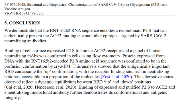
One of the most recent documents published by the FDA saved as 125742_S1_M4_4.2.1 vr vtr 10741.pdf, confirms the use of Graphene Oxide in the manufacturing process of the Pfizer Covid-19 vaccine.
The document is a description of a study carried out by Pfizer between April 7th 2020 and 19th August 2020, with the objective being “to express and characterize the vaccine antigen encoded by BNT162b2.”
In layman’s terms, the study was conducted to determine how the vaccine works. The study found that the vaccine used mRNA to instruct your cells to produce a protein (called P2 S), which is the Spike protein of the alleged Covd-19 virus.
The millions of spike proteins then bind to a receptor called ACE2 on the surface of your cells, inducing an immune system response.
But what is most interesting about the study is that it confirms on page 7 that reduced Graphene Oxide is required to manufacture the Pfizer Covid-19 vaccine because it is needed as a base for the lipid nanoparticles.
Pfizer states on page 7 of the study in section 3.4 the following –
This is most peculiar because medicine regulators with the help of the Mainstream Media, have denied for months on end that Graphene Oxide is an ingredient of the Covid-19 vaccine. They’ve been able to say this because those who’ve proven and speculated Graphene Oxide is in the Pfizer Covid19 injection have been asking the wrong question.
What everyone should have been asking is, ‘is Graphene Oxide used in the manufacturing process of the Pfizer Covid vaccine?’
Because as this document, which the FDA attempted to keep confidential and sealed the 75 years, shows, Graphene Oxide is indeed used in the manufacturing process of the vaccine because it is vital in helping to make the vaccine’s lipid nanoparticles stable.
Therefore, trace amounts or large amounts, depending on the batch, of reduced Graphene Oxide inevitably make their way into the Pfizer Covid-19 injections.
What are Lipid Nanoparticles?
The Pfizer Covid-19 vaccine uses tiny particles called lipid nanoparticles to deliver the vaccine’s genetic material (called messenger RNA, or mRNA) into cells in the body. These lipid nanoparticles are like tiny “bubbles” made up of fats and other molecules that can surround and protect the mRNA until it reaches its destination inside the cells.
The mRNA in the vaccine provides instructions to the cells to produce a protein (called spike protein) that is found on the surface of the Covid-19 virus. When the immune system detects this spike protein, it can recognize it as foreign and mount an immune response against it,
Furthermore, it has been discovered that two of the lipids in Pfizer Covid-19 vaccines are ALC-0159 and ALC-315, as confirmed by the assessment report of the Pfizer Covid-19 vaccine published by the European Medicines Agency.
Graphene Oxide is a tiny particle that is made up of carbon and oxygen atoms. It’s really small – so small that you can’t see it with your eyes. But even though it’s small, it can be dangerous.
It is known to be toxic to some cells, and research has shown that it can cause inflammation and damage to the lungs when inhaled.
In addition, Graphene Oxide is able to cross the blood-brain barrier, which is a protective barrier that prevents harmful substances from entering the brain. This could potentially lead to neurological problems.
Graphene Oxide is detected in the immune system as if it were a pathogen. Once injected it has an affinity for the central nervous system, potentially causing paralysis, strokes and alteration of the nervous system.
Furthermore, the long-term effects of Graphene Oxide exposure are not yet known. There is very little research on the long-term effects of Graphene Oxide exposure in humans, which means we don’t know what the potential risks are.
But thanks to the administration of the Pfizer COVID vaccine to millions of people, numerous times, we are finding out as the days pass. And sadly, both the short-term and long-term effects do not look good.
Further Evidence, Other Undeclared Substances & Deadly Consequences
Graphene Oxide is not the only toxic substance that the public should be concerned about. Because scientists have found Nanotech alongside Graphene Oxide when they have previously examined samples of Covid-19 injections.
Nanoscience and nanotechnology involve the ability to see and control individual atoms and molecules. Everything on Earth is made up of atoms—the food we eat, the clothes we wear, the buildings and houses we live in, and our own bodies.
But something as small as an atom is impossible to see with the naked eye. In fact, it’s impossible to see with the microscopes typically used in high school science classes. The microscopes needed to see things at the nanoscale were invented in the early 1980s.
Once scientists had the right tools, such as the scanning tunnelling microscope (STM) and the atomic force microscope (AFM), the age of nanotechnology was born.
And scientists from Spain, have declared that nanotechnology, which has the ability to control atoms in your body, has been found in all of the Covid-19 injections alongside Graphene Oxide. https://odysee.com/$/embed/@StopTheCrime:d/Breaking-News-SHOCKING—Here-is-What-Really-is-in-the-Vaccines:d?r=7w5QuSaWFRbu1bEVToV9pb1Zdn8mheAq
According to the Spanish scientists who examined the Covid-19 injections, Graphene Oxide has the potential to cause strange blood clots. This may explain why it has been proven that Covid-19 injections increase the risk of suffering thrombosis with thrombocytopenia.
But it is not just scientists from Spain making these claims. Numerous scientists around the world have published findings where they allege they have discovered both nanotechnology and Graphene Oxide in the Covid-19 injections.
After reviewing electron microscope images of elements contained in the Covid Pfizer and Moderna injections, Dr Daniel Nagase, a Canadian emergency room doctor, revealed that, strangely, the contents of the Pfizer and Moderna “vaccines” show no signs of biological material, including mRNA or DNA. (Read more here).
Dr Nagase’s research group looked at Moderna and Pfizer samples under a regular microscope. Although there were a lot of very interesting images, they were unable to be conclusive about what exactly they were seeing. So, they used an electron microscope to determine what elements the “vaccines” contained.
Here are some of the images of what they found –
Found in a Moderna Covid “vaccine” sample
Dr Nagase examined a “ball with the legs growing out of it” found inside a Moderna sample and had this to say –
“This shape, this ball with the legs growing out of it, for some reason has aluminium in it. And I can say with certainty that this isn’t a mould spore or some other type of biological contamination, because the only thing in it is carbon, oxygen, and no signs of nitrogen, no signs of phosphorus, which would indicate something biological of origin. So, this thing that’s growing is non-biological.”
Dr Nagase and the researchers also discovered an unusual element from the lanthanide series – thulium – in a fibre-like structure found in a Pfizer sample –
Found in a Pfizer Covid “vaccine” sample
Dr. Nagase and the researchers found a variety of shapes and structures inside the “vaccine” samples they tested – crystals, chips, strands, bulbs, spheres, fibres and balls with legs growing out of them – “we have polymorphic, which is many different forms,” he said.
“They all seem to be made predominantly out of carbon and oxygen and they were in both the Moderna and Pfizer samples, and they seem to be in fibre forms. In the Moderna sample, the carbon-oxygen structures seem to be taking nanosphere forms and crystalline forms. And in the Pfizer sample … seem to only be forming fibres and crystals.
In a presentation to the Chilean radio station El Mirador del Gallo, Argentine doctor Martín Monteverde presented the analyses carried out by Corona2Inspect researchers on the microtechnology found in the Pfizer Covid-19 mRNA vaccine.
Argentina’s Dr Monteverde and other researchers carried out microscopic analyses of a vial of the Pfizer vaccine alongside four other Covid-19 “vaccine” types. He then sent these images to Corona2Inspect for further analysis. Corona2Inspect returned the images with their comments identifying what objects the images were showing.
You can watch a video of Dr Monteverde’s teams findings below – https://www.bitchute.com/embed/rp5ZyrmMLJQv/
Argentina’s Dr Patricia Aprea, Director of Evaluation and Control of the ANMAT, also accidentally admitted AstraZeneca’s Viral Vector Covid-19 injection also contains Graphene during a legal case regarding a death post-Covid injection.
You can read the document where ANMAT recognised that Covid-19 vaccines contain Graphene Oxide HERE in (Spanish) or below, translated into English using Google –
Dr Philippe van Welbergen, Medical Director of Biomedical Clinics, was one of the first to warn the public of the damage being caused to people’s blood by Covid injections by releasing images of blood samples under the microscope.
In a set of slides of blood samples taken from both “vaccinated” and unvaccinated people, Dr Philippe van Welbergen demonstrated that the Graphene Oxide, contained in the Pfizer Covid-19 vaccines being injected into people by amateurs and volunteers with no adequate training, is organising and growing into larger fibres and structures, gaining magnetic properties or an electrical charge and the fibres are showing indications of more complex structures with striations.
At the beginning of July 2021, Dr Philippe, was interviewed and he explained that when his patients started complaining about chronic fatigue, dizziness, memory loss, and even sometimes paralysis and late onset of heavy menstruation (women in their 60s upwards), he took blood samples.
Their blood had unusual tube-like structures, some particles which lit up and many damaged cells.
Few healthy cells were visible. Until three months earlier, he had never seen these formations in blood.
We now know these tube-like structures are Graphene Oxide.
He also demonstrated that “shards” of Graphene Oxide are being transmitted from the Covid-19 vaccinated to vaccine-free or unvaccinated people, sadly destroying their red blood cells and causing blood clots. (Read more here).
Below is an image of typical healthy red blood cells as seen with a microscope, what blood should look like. There is no coagulation or foreign objects in it.
Sadly fibres of this size are capable of blocking capillaries.
You can also see that the Graphene fibres are hollow and have swallowed up some red blood cells.
In December 2021, a British medical practitioner offered to assist in an investigation to ascertain whether the results discovered by Dr. Andreas Noack, a German chemist, and Dr. Pablo Campra, of the University of Almeria in Spain, could be replicated in the UK and also to examine the Covid-19 injection vials for toxins or unexpected contents.
The medical practitioner seized an injection vial from the fridge housed in the surgery where she works and handed it to an independent investigator.
A UK laboratory analysed the sample using Raman Spectroscopy and found Graphene, SP3 carbon, iron oxide, carbon derivatives and glass shards.
The first sample that was evaluated was the Moderna 01 which was examined by Raman spectroscopy. The investigation clearly showed that all the inclusions within the vaccine have a strong carbon signal with confirmed graphene compositions of some representative forms.
Two clear signals were obtained from two objects. The flat ribbon-like inclusions exhibited clear Graphene spectra integrated with the spectrum of glycol and other minor compounds. The other clear signal was obtained from a calcite microcrystalline form and Carbon composite forms also had a clear Graphene signal.
You can read a copy of the document encompassing a case briefing, the UNIT report and a summary of the toxicity of Graphene nanoparticles on UK Citizen 2021’s website HERE.
The 48-page UNIT report, ‘Qualitative Evaluation of Inclusions in Moderna, AstraZeneca and Pfizer Covid-19 vaccines’, begins on page 12 of the document.
An Open Access review highlighting the toxicity of the graphene family nanoparticles can be viewed here.
Nanotechnology and Graphene have also been found in Pfizer’s Comirnaty “vaccines” by scientists in New Zealand. (Read more here).
At the end of January 2022, Sue Grey, co-leader of the Outdoors and Freedom Party, and Dr Matt Shelton from New Zealand Doctors Speaking Out With Science (“NZDSOS”) put the Health Select Committee on notice that serious contamination of the Pfizer vaccine has been uncovered and they needed to act immediately to stop the injection campaign.
Dr Shelton came forward to disclose the discovery of formations of nano-particles found by New Zealand scientists using specialised microscopic techniques.
None of the experts consulted had ever seen anything like this before, and none of these contaminants are listed as approved ingredients.
You can read the full account, with additional images and videos, HERE. But below is a snapshot of what one New Zealand scientist found.
The image below was taken from one drop of New Zealand’s Pfizer Cominarty “vaccine” under a cover slip, after it was inadvertently heated lightly, and viewed the same day through dark field microscopy at low magnification, projected onto a TV monitor.
The following images were taken after a new computer with improved graphics was purchased alongside new software for the camera –
They lied to you
Despite repeated assurances from authorities and mainstream media that the Covid-19 vaccines are safe and effective, evidence has emerged time and time again that proves they have not been telling the whole truth.
The use of Graphene Oxide in the Pfizer Covid-19 vaccine has been a source of controversy and concern from the outset, with many individuals claiming that regulators and media outlets were deliberately misleading the public about its inclusion.
Despite initial denials, the documents released by the FDA, which they were forced to publish by order of the Federal Court in the USA. have confirmed the use of Graphene Oxide in the manufacturing process of the Pfizer vaccine, raising questions about who we can trust.
This revelation should cause widespread alarm and will likely fuel suspicion about the true intentions of those in charge of public health.
When I discovered this study several years ago and wrote the following extensive piece on it, the study was a bolt from the blue, a complete devastating shocker.
It still is.
It is more than enough to topple the whole vaccine empire.
Honoring the work of the study co-author, Dr. Antonietta Gatti, Catherine Austin Fitts wrote, “Not long after the publication of this revolutionary study, tax authorities raided and investigated Dr. Gatti’s and [her husband] Dr. Montanari’s laboratory and private home—an all too usual method of intimidation.”
THAT was the “scientific follow-up.”
In a nutshell, Dr. Gatti’s 2017 study showed an incredible amount of contamination, in a whole host of traditional vaccines. The contamination was in the form of tiny nanoparticles, mostly metallic, and obviously highly harmful and dangerous.
Before reading my summary and analysis of that study—here is an updated communication from Dr. Gatti I received a few days ago. It describes, in a stark and disturbing fashion, what has been happening to her, her work, and her laboratory. This is chilling:
“At the end of last year, our laboratory no longer had the financial capacity to continue its research. The proceeds from the few analyzes requested by private individuals yielded enormously less than what the research cost us. Then, there were two possibilities: close everything or set up a foundation by giving away everything that belonged to us, hoping to find some sponsors. After all, all initiatives, even the most bizarre, find someone willing to contribute financially. Why not a foundation that does fundamental research on health? So, we opted for the latter choice, and the Nanodiagnostics Foundation was born.”
“But, after almost a year, not a cent has arrived. In short, no company, no private citizen, no institution is willing to contribute.”
“Many people continue to demand results and ask questions to which they have no answers from the institutions or their doctors, but, if it is a question of parting from some money, the silence is absolute.”
“It is clear that our work is a threat to billion-dollar businesses that are not exactly clear, at least for most people. For this reason, the most absurd and incredible slanders are invented to our detriment.
Not being able to dispute our scientific results, there are those who publish, usually anonymously, that we earn enormous sums of money, even giving the impression that the Foundation belongs to us, when it should be known that foundations do not belong to anyone, and no one can profit from them. And this is when we have donated everything that belonged to us, and we work for free.”
“Another tactic is trying to isolate and discredit us with lies. What the University of Bologna did a few days ago, the university where I graduated, then specialized and taught, is a small example.”
“A few months ago, that university asked us if we were willing to accept [a] student… who would prepare her graduation thesis with us. We agreed and agreed with the student on how to proceed. A few months passed, then, a couple of weeks ago, when the University authorities realized that the student would work with us, they sent us a message of a few lines in which they informed us that what we do (and which I had taught at that university) was of no interest to them (which, in a way, is true, although very far from the mission of the University). Needless to say, my letter to the Rector asking for explanations remained unanswered.”
“And it is also useless to say how difficult it is to publish the results that we continue to obtain, and which are not liked by those who financially maintain the medical journals, on whose scientific nature I prefer not to comment. For twice the Editor after the publication of an article (on vaccines and on SIDS) asked to retreat [sic] them. Only the work of the Robert Kennedy Jr lawyers stopped the request.”
“[Paper:] Novel chemical-physical autopsy investigation in sudden infant death and sudden intrauterine unexplained death syndromes” (click here)
“Just for your information, in spite of all difficulties, we are now dealing with very critical topics: spontaneously aborted babies, analysis of the brains of infants who died in cots (Sudden Infant Death Syndrome, aka SIDS), analysis of what falls from the sky (e.g., recently hail never seen before), food, etc. All this can only be fought with personal discredit.”
“We haven’t had any visits from the regime for a long time. For them it is enough to monitor our computers and phones. The rest is done by ‘volunteers’. As for other scientists, no one deals with our topics in full. It must be realized that doing so represents a risk that is obviously preferable not to take.”
“As long as we can manage, we will continue to work. If, however, no sponsor materializes (idle chatter and empty promises are not only useless: they are a waste of time,) we will have no other option than to declare defeat, a defeat that belongs to the whole world and, above all, to the children who do not deserve the fate they are suffering.”
“…I give some details of our Foundation Nanodiagnostics (click here)…”
IF YOU CAN, PLEASE DONATE TO Dr. Gatti’s vital work at the above website.
Here is my original article on Dr. Gatti’s vaccine-contamination study:
Dangerous nano-particles contaminating many vaccines: groundbreaking study
“The Lung,” Second Edition: “Nanoparticles [are] comparable in size to subcellular structures…enabling their ready incorporation into biological systems.”
A 2017 study of 44 types of 15 traditional vaccines, manufactured by leading global companies, has uncovered a very troubling and previously unreported fact:
The vaccines are heavily contaminated with a variety of nanoparticles.
Many of the particles are metals.
We’re talking about traditional vaccines, such as HPV, flu, Swine Flu, Hepatitis B, MMR, DPT, tetanus, etc.
To begin to understand some of the destructive effects of contaminating nanoparticles in vaccines, here is the groundbreaking 2017 study:International Journal of Vaccines & Vaccination Volume 4 Issue 1 January 23 2017 New Quality-Control Investigations on Vaccines: Micro- and Nanocontamination Antonietta M Gatti and Stefano Montanari (Paper archived here and here)
“The analyses carried out show that in all samples checked vaccines contain non biocompatible and bio-persistent foreign bodies which are not declared by the Producers, against which the body reacts in any case. This new investigation represents a new quality control that can be adopted to assess the safety of a vaccine. Our hypothesis is that this contamination is unintentional, since it is probably due to polluted components or procedures of industrial processes (e.g. filtrations) used to produce vaccines…”
Are the study authors leaving the door open to the possibility that the contamination is intentional?
“The quantity of foreign bodies detected and, in some cases, their unusual chemical compositions baffled us. The inorganic particles identified are neither biocompatible nor biodegradable, that means that they are biopersistent and can induce effects that can become evident either immediately close to injection time or after a certain time from administration. It is important to remember that particles (crystals and not molecules) are bodies foreign to the organism and they behave as such. More in particular, their toxicity is in some respects different from that of the chemical elements composing them, adding to that toxicity…they induce an inflammatory reaction.”
“After being injected, those microparticles, nanoparticles and aggregates can stay around the injection site forming swellings and granulomas…But they can also be carried by the blood circulation, escaping any attempt to guess what will be their final destination…As happens with all foreign bodies, particularly that small, they induce an inflammatory reaction that is chronic because most of those particles cannot be degraded. Furthermore, the protein-corona effect…due to a nano-bio-interaction…can produce organic/inorganic composite particles capable of stimulating the immune system in an undesirable way…It is impossible not to add that particles the size often observed in vaccines can enter cell nuclei and interact with the DNA…”
“In some cases, e.g. as occurs with Iron and some Iron alloys, they can corrode and the corrosion products exert a toxicity affecting the tissues…”
“Given the contaminations we observed in all samples of human-use vaccines, adverse effects after the injection of those vaccines are possible and credible and have the character of randomness, since they depend on where the contaminants are carried by the blood circulation. It is only obvious that similar quantities of these foreign bodies can have a more serious impact on very small organisms like those of children. Their presence in the muscles…could heavily impair the muscle functionality…”
“We come across particles with chemical compositions, similar to those found in the vaccines we analyzed, when we study cases of environmental contamination caused by different pollution sources. In most circumstances, the combinations detected are very odd as they have no technical use, cannot be found in any material handbook and look like the result of the random formation occurring, for example, when waste is burnt. In any case, whatever their origin, they should not be present in any injectable medicament, let alone in vaccines, more in particular those meant for infants.”
This 2017 study opens up a whole new field: the investigation of nanoparticles in vaccines where none were expected.
Such particles are not medicine in any sense of the word.
Many legal and scientific “experts” assert the State has a right to mandate vaccines and force them on the population. But these contaminating nanoparticles are not vaccines or medicines. Only a lunatic would defend the right of the State to inject them.
Here is another section from the 2017 study. Trade names of vaccines, and compositions of the nanoparticle contaminants are indicated. Take a deep breath and buckle up:
“…further presence of micro-, sub-micro- and nanosized, inorganic, foreign bodies (ranging from 100nm to about ten microns) was identified in all cases [all 44 vaccines], whose presence was not declared in the leaflets delivered in the package of the product…”
“…single particles, cluster of micro- and nanoparticles (less than 100nm) and aggregates…debris of Aluminum, Silicon, Magnesium and Titanium; of Iron, Chromium, Silicon and Calcium particles…arranged in a cluster, and Aluminum-Copper debris…in an aggregate.”
“…the particles are surrounded and embedded in a biological substrate. In all the samples analyzed, we identified particles containing: Lead (Typhym, Cervarix, Agrippal S1, Meningitec, Gardasil) or stainless steel (Mencevax, Infarix Hexa, Cervarix. Anatetall, Focetria, Agrippal S1, Menveo, Prevenar 13, Meningitec, Vaxigrip, Stamaril Pasteur, Repevax and MMRvaxPro).”
“…particles of Tungsten identified in drops of Prevenar and Infarix (Aluminum, Tungsten, Calcium chloride).”
“…singular debris found in Repevax (Silicon, Gold, Silver) and Gardasil (Zirconium).”
“Some metallic particles made of Tungsten or stainless steel were also identified. Other particles containing Zirconium, Hafnium, Strontium and Aluminum (Vivotif, Meningetec); Tungsten, Nickel, Iron (Priorix, Meningetec); Antimony (Menjugate kit); Chromium (Meningetec); Gold or Gold, Zinc (Infarix Hexa, Repevax), or Platinum, Silver, Bismuth, Iron, Chromium (MMRvaxPro) or Lead,Bismuth (Gardasil) or Cerium (Agrippal S1) were also found. The only Tungsten appears in 8/44 vaccines, while Chromium (alone or in alloy with Iron and Nickel) in 25/44. The investigations revealed that some particles are embedded in a biological substrate, probably proteins, endo-toxins and residues of bacteria. As soon as a particle comes in contact with proteic fluids, a nano-bio-interaction…occurs and a ‘protein corona’ is formed…The nano-bio-interaction generates a bigger-sized compound that is not biodegradable and can induce adverse effects, since it is not recognized as self by the body.”
“…examples of these nano-bio-interactions. Aggregates can be seen (stable composite entities) containing particles of Lead in Meningitec… of stainless steel (Iron, Chromium and Nickel…) and of Copper, Zinc and Lead in Cervarix…Similar aggregates, though in different situations (patients suffering from leukemia or cryoglobulinemia), have already been described in literature.”
I’m sure you’ve read official assurances that vaccine-manufacturing problems are “rare.” You can file those pronouncements along with other medical lies.
“I’d like the heavy metal sandwich on rye, please. And instead of serving it on a plate, can you inject it?”
Several vital questions demanding answers spring from the findings of this 2017 study:
Are some of these nanoparticles intentionally placed in vaccines?
Does the standard manufacturing process for traditional vaccines INEVITABLY lead to dangerous and destructive nano-contamination?
New nano-technology is already being employed to create several vaccines—supposedly “improving effectiveness.” In fact, the RNA COVID-19 vaccine are a nano-type. Does this manufacturing process carry with it the unavoidable effect of unleashing a hurricane of nanoparticle contaminants?
How many cases of childhood brain damage and autism can be laid at the door of nanoparticle contamination?
And finally, where are these contaminated vaccines manufactured? The above study did not attempt to discover this. It was outside the scope of the research. It’s common knowledge that, for example, in the case of the US, vaccines or their components, are, in many instances, not produced domestically. Where does this put control of safety? In, say, China, where there have been numerous pharmaceutical scandals connected to contamination of products?
The vaccine establishment does not show the slightest interest in answering any of these questions. They are busy pretending the questions don’t exist.
In case you are unable to read them on above links, download from below, both books in PDF. Plus more books on poisonous needles aka vaccines… Uh spare me Fuck them vaccines!
Are you getting bored of listening to the exact same song and dance numbers over and over again? Are the old “viral” bands just not doing it for you anymore? Are you looking for something new and mysterious to come along in order to spice things up a bit and reignite the dwindling levels of fear? If so, then you are in luck as there is a brand new “viral” sensation headed your way!
Introducing Disease X!
“An old adage says, “Prevention is better than cure.” Nothing exemplifies this idea better than “Disease X.” According to the World Health Organization (WHO), “Disease X represents the knowledge that a serious international epidemic could be caused by a pathogen currently unknown to cause human disease.”1
Richard Hatchett, chief executive officer (CEO) of the Coalition for Epidemic Preparedness Innovations (CEPI), said about Disease X, “It might sound like science fiction, but Disease X is something we must prepare for.”2 In a list of diseases that the WHO considers high priority in terms of research and development, Disease X occupies a spot among diseases such as Ebola, Zika, and coronavirus disease 2019 (COVID-19).1Unexpected outbreaks of infectious disease (Disease X) have repeatedly rocked the medical confidence and have taken the medical world by surprise.3
Some experts have even commented that COVID-19, caused by severe acute respiratory coronavirus virus 2 (SARS-CoV-2), met the standards to be considered the first Disease X,4 while some authors have called Zika a Disease X.5However, one unfortunate possibility is that COVID-19 and other recent pandemics might have been milder versions of what will eventually be the most prominent Disease X.
Disease X is supposed to be caused by a “pathogen X.” Such a pathogen is expected to be a zoonosis, most likely an RNA virus, emerging from an area where the right mix of risk factors highly promotes the risk for sustained transmission.6
Bloody quacks should pay attention ! As most of them the so called @awakened@ were fast asleep and only started scratching the surface once they saw that their “students” were getting upset about their willful ignorance in this regard ! What a pity such “DOCTORS” are – Shame on you ! Yeah you know I… Continue reading Independent researchers find no evidence of mRNA in the COVID injection.
The headline of this article has become a battle cry among some “alternative journalists,” activists, lawyers, and doctors. As my readers know, I’ve devoted considerable space, over the past two years, to presenting evidence that SARS-CoV-2 is a scientific fairy tale, a con, and the virus doesn’t exist. So when I hear this battle cry,… Continue reading “Stop arguing about the existence of the virus” by Jon Rappoport
In interviews with The Defender, pilots injured by COVID-19 vaccines said despite a “culture of fear and intimidation” they are compelled to speak out against vaccine mandates that rob pilots of their careers — and in some cases their lives. As a commercial pilot, Bob Snow had long looked forward to seeing his daughter follow… Continue reading Pilots Injured by COVID Vaccines Speak Out: ‘I Will Probably Never Fly Again’